
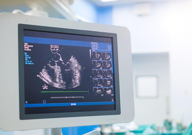
An echocardiogram, often referred to as an “echo,” is a diagnostic test that uses sound waves to create detailed images of the heart. This non-invasive imaging test helps healthcare professionals assess the structure and function of the heart, providing valuable information about the heart’s chambers, valves, and overall performance.
Types of Echocardiograms
⦁ Transthoracic Echocardiogram (TTE):
- Procedure: A handheld device called a transducer is placed on the chest to capture images of the heart through the chest wall.
- Common Use: General assessment of heart structure and function.
⦁ Transesophageal Echocardiogram (TEE):
- Procedure: A specialized transducer is passed into the esophagus to obtain images from a closer proximity to the heart.
- Common Use: Provides more detailed images, often used for closer examination of the heart valves and chambers.
⦁ Stress Echocardiogram:
- Procedure: Combines an echocardiogram with physical stress (exercise or medication) to evaluate heart function under increased workload.
- Common Use: Assessing coronary artery disease and evaluating exercise-induced changes in heart function.
What Echocardiograms Can Assess
⦁ Heart Chamber Size:
- Evaluate the size of the heart chambers to identify any enlargement or abnormalities.
⦁ Valve Function:
- Assess the movement and function of heart valves, checking for conditions such as valve stenosis or regurgitation.
⦁ Ejection Fraction:
- Measure the percentage of blood pumped out of the heart with each contraction, helping to assess overall heart function.
⦁ Blood Flow:
- Examine the flow of blood through the heart and its major vessels.
⦁ Structural Abnormalities:
- Identify any structural abnormalities, such as tumors or blood clots, within the heart.
Preparation for an Echocardiogram
⦁ Clothing:
- Wear comfortable clothing, and in some cases, a hospital gown may be provided.
⦁ Medication:
- Follow any specific instructions regarding the use of medications on the day of the test.
⦁ Fasting:
- In some cases, fasting may be required before the test.
How the Test is Performed
⦁ TTE:
- A gel is applied to the chest, and the transducer is moved across different areas to obtain images.
⦁ TEE:
- A specialized transducer is inserted through the mouth into the esophagus, providing clearer images of the heart.
⦁ Stress Echocardiogram:
- Images are obtained before, during, and after stress (exercise or medication-induced), allowing for a comparison of heart function under different conditions.
Importance and Clinical Use:
⦁ Diagnosis and Monitoring:
- Diagnose various heart conditions, including heart valve disorders, heart failure, and congenital heart defects.
⦁ Treatment Planning:
- Aid in the planning of treatment strategies for heart conditions.
⦁ Post-Cardiac Procedures:
- Assess the success of certain cardiac procedures, such as valve replacements or repairs.
⦁ Cardiovascular Risk Assessment:
- Evaluate cardiovascular risk factors and assess overall heart health.
Benefits
⦁ Non-Invasive:
- Echocardiograms are non-invasive and do not involve radiation.
⦁ Real-Time Imaging:
- Provide real-time images of the heart’s structure and function.
⦁ Versatility:
- Useful for a wide range of cardiac conditions and assessments.
Limitations
⦁ Operator Dependency:
- Image quality can depend on the skill and experience of the operator.
⦁ Limited Views:
- In certain patients, such as those with obesity or lung disease, obtaining clear images may be challenging.
⦁ Not Always Diagnostic:
- In some cases, additional tests may be needed for a comprehensive evaluation.
An echocardiogram is a valuable tool in cardiology, offering insights into the structure and function of the heart. It plays a crucial role in diagnosing and monitoring various cardiovascular conditions, helping healthcare professionals make informed decisions about treatment and patient care.
Stress Test
Stress tests are also called exercise stress tests as it examines how your heart handles physical exercise. You will be connected to an electrocardiogram (ECG) to monitor your heart's electrical rhythms as you run a treadmill or work a stationary bicycle. By making your heart work steadily harder and faster, the stress test can unveil blood flow problems in your heart.
Doctors conduct stress tests if you have symptoms like chest pain, irregular heartbeat, or determine your health level. The stress test is necessary as it helps to:
- Diagnose coronary artery disease
- Diagnose arrhythmia
- Evaluate the supply of blood to your heart
- Determine your heart's health if you are at risk for any heart disease
- Find a safe level of exercise
Stress tests are usually safe, and there is no need to worry as trained medical professionals will closely monitor you. However, even if the probability of risks is low, some complications that may occur are:
⦁ Dizziness
⦁ Chest pain
⦁ Nausea
⦁ Abnormal heart rhythm
Stress tests may not be appropriate for everyone. Your physician will decide if the test is necessary for you based on your symptoms, medical history, age, sex, level of physical activity, and other risk factors.
Your physician will inform you on how to prepare for the stress test. It may include instructions like:
⦁ Avoid eating or drinking (especially caffeinated products) several hours prior to the test.
⦁ Avoid smoking or tobacco products
⦁ You may need to stop taking specific medications that may hinder the test if advised by your physician
⦁ Wear loose-fitting or comfortable clothes and walking shoes for the test